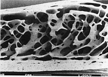
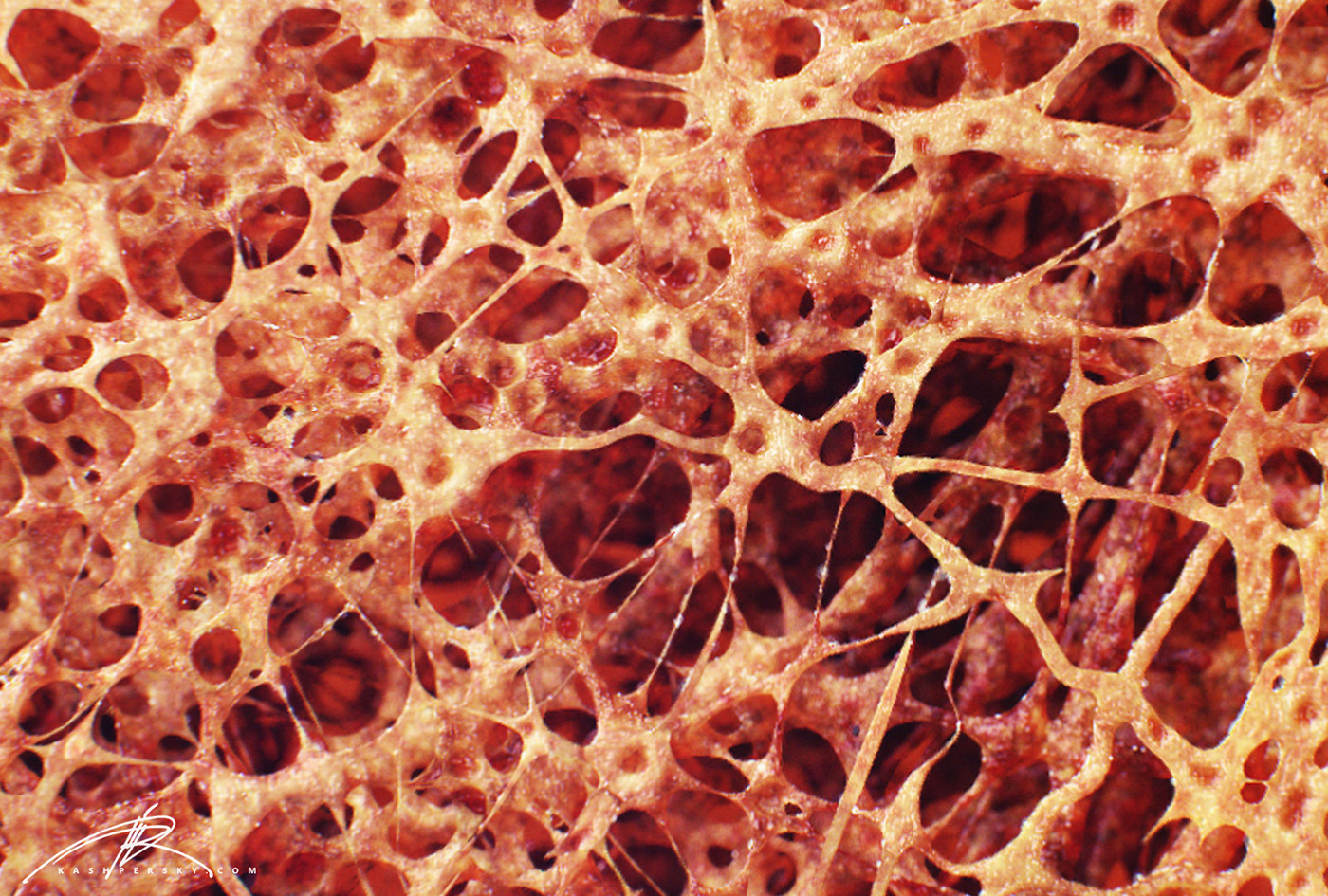
Bone Cross Section - Osteoporosis Cross Section Image Osteoporosis Bone Stock Vector 776790541 - Shutterstock : An outer 'fibrous layer' containing mainly fibroblasts, and an inner 'cambium layer' containing progenitor cells.. While it is not as hard as compact bone, spongy bone plays an important role of protecting the marrow where blood cells are produced. Compact bone, dense bone in which the bony matrix is solidly filled with organic ground substance and inorganic salts, leaving only tiny spaces that contain the osteocytes, or bone cells. For example, if i missed labeling anything, or any parts of the bone are missing. This is a short tutorial using blender 2.8 that shows how to create a bone cross section and using images to create the textures.hope you enjoy and please su. A demineralized bone preparation is one in which the the mineral has been removed, leaving behind the organic components of the tissue.
Vector illustration scheme of bone cross section. This is known as the periosteum. Bone structure right foot 12 photos of the bone structure right foot bone structure in. Browse 53 bone marrow cross section stock photos and images available, or search for bone cross section or bone cells to find more great stock photos and pictures. I am not an expert on this subject, so i was wondering if anyone could put their input on this image.
Bone Fracture Healing Explained - Fractures & Broken Bones from www.physioroom.com A) the elastic modulus b) the yield strength c) the ultimate strength. Photomechanical print page item number: Compact bone is the outer layer and the spongy bone forms the inner layer. Browse 9,121 bone cross section stock photos and images available, or search for bone marrow or bone structure to find more great stock photos and pictures. I am not an expert on this subject, so i was wondering if anyone could put their input on this image. This is known as the periosteum. Slides have to be made this way because the matrix of bone is too hard to The remainder is spongelike cancellous bone.
Can you identify the concentric lamellae, central canal and the lacunae.
You may do so in any reasonable manner, but not in any way. Compact bone is the outer layer and the spongy bone forms the inner layer. This file is licensed under the creative commons attribution 3.0 unported license.: This slide contained a cross section of a very small bone, and you are looking at the entire thickness of the shaft of the bone. This photo shows a cross section through bone. Bone structure right foot 12 photos of the bone structure right foot bone structure in. For example, if i missed labeling anything, or any parts of the bone are missing. Beautiful tooth cross section illustration, deep blue background and sparkling lights around. A) the elastic modulus b) the yield strength c) the ultimate strength. Human bone, cross section diagram of femur showing osteon, veins, marrow. And the bone marrow.the femur is the thigh bone, the longest bone in the body. Bone cross section 3d, formats max, 3d anatomy bone cell cross cutaway detailed, ready for 3d animation and other 3d projects Slides have to be made this way because the matrix of bone is too hard to
You may do so in any reasonable manner, but not in any way. Related posts of cross section of human bone diagram muscles and bones of the human body. If we were to cut the femur bone in half, we would see that it contains various layers. As the names suggest compact bone looks compact and the spongy bone looks like sponges. Cross‐sectional area is derived from the integral of the bone mass profile across the narrow region.
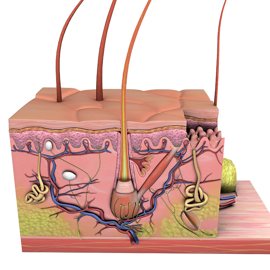
Anatomy Of Skin, Cross Section On White Photograph by Hank Grebe from images.fineartamerica.com Bone structure right foot 12 photos of the bone structure right foot bone structure in. The figure below shows the stress strain diagram of a cortical bone, with initially rectangular cross section of 15 x 20 mm², and length of 10 cm. Photomechanical print page item number: Compact bones make up 80 percent of the human skeleton; If we were to cut the femur bone in half, we would see that it contains various layers. Fetal leg, cross section, masson stain, 40x (spongy bone, appositional bone growth on surface of long bone). Related posts of cross section of human bone diagram muscles and bones of the human body. After a fracture, woven bone forms initially and is gradually replaced by lamellar bone during a process known as bony substitution.
Slides have to be made this way because the matrix of bone is too hard to
A demineralized bone preparation is one in which the the mineral has been removed, leaving behind the organic components of the tissue. Bone cross section 3d, formats max, 3d anatomy bone cell cross cutaway detailed, ready for 3d animation and other 3d projects This file is licensed under the creative commons attribution 3.0 unported license.: Cross‐sectional area is derived from the integral of the bone mass profile across the narrow region. While it is not as hard as compact bone, spongy bone plays an important role of protecting the marrow where blood cells are produced. Obtain a demineralized compact bone preparation (in cross section), preferably from the diaphysis of a long bone, and prepare to examine it microscopically. A) the elastic modulus b) the yield strength c) the ultimate strength. It consists of two layers; Related posts of cross section of a long bone bone structure right foot. After a fracture, woven bone forms initially and is gradually replaced by lamellar bone during a process known as bony substitution. Browse 9,121 bone cross section stock photos and images available, or search for bone marrow or bone structure to find more great stock photos and pictures. Muscles and bones of the human body 12 photos of the muscles and bones of the human body anatomy bones of the human body quiz, major muscles and bones in the human body, muscles and bones in the human body, number of muscles and bones in the human body. Diagram with articular cartilage, marrow, spongy bone, medullary cavity, endosteum, diaphysis, and periosteum.
After a fracture, woven bone forms initially and is gradually replaced by lamellar bone during a process known as bony substitution. Two types of bone tissues in cross section of a long bone : This is a high power photo of a single haversian system. Related posts of cross section of human bone diagram muscles and bones of the human body. Compact bone is very different from the other tissues you have seen.
"Bone Cross Section" for Radius Digital Science on Behance from mir-s3-cdn-cf.behance.net As the names suggest compact bone looks compact and the spongy bone looks like sponges. Photomechanical print page item number: A) the elastic modulus b) the yield strength c) the ultimate strength. It consists of two layers; Compact bones make up 80 percent of the human skeleton; Beautiful tooth cross section illustration, deep blue background and sparkling lights around. Related posts of cross section of human bone diagram muscles and bones of the human body. A demineralized bone preparation is one in which the the mineral has been removed, leaving behind the organic components of the tissue.
Related posts of cross section of human bone diagram muscles and bones of the human body.
As the names suggest compact bone looks compact and the spongy bone looks like sponges. Can you identify the primary and secondary haversian systems, central canals and bone lamellae? Two types of bone tissues in cross section of a long bone : They are obtained by taking imaginary slices perpendicular to the main axis of organs, vessels, nerves, bones, soft tissue, or even the entire human body. Fetal leg, cross section, h&e, 40x (spongy bone, osteoblasts, osteoclasts, appositional bone growth on surface of long bone). Compact bone, dense bone in which the bony matrix is solidly filled with organic ground substance and inorganic salts, leaving only tiny spaces that contain the osteocytes, or bone cells. This photo shows a cross section through bone. The remainder is spongelike cancellous bone. And the bone marrow.the femur is the thigh bone, the longest bone in the body. Explaned distal and proximal epiphysis. To the left is muscle tissue, and to the right Bone structure right foot 12 photos of the bone structure right foot bone structure in. The compact bone is made up of osteon.
0 Comments:
Posting Komentar